How 3D Digital Scanning Improves Precision in Oral and Facial Surgery
15 December 2025

Oral and facial surgery requires an exceptional level of precision to ensure optimal outcomes for patients. Traditional methods, while effective in many cases, often rely on manual impressions and estimations that can introduce minor inaccuracies. With the advent of modern dental technology, 3D digital scanning oral surgery in Pinole, CA has become a game-changer in delivering highly accurate results. By integrating 3D scanning technology, surgeons can now achieve precision and predictability previously unattainable in oral and facial procedures.
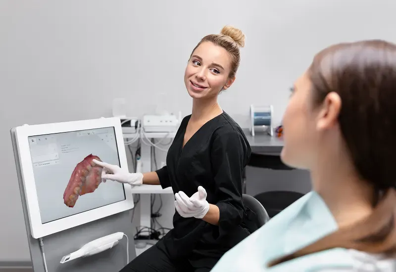
At the heart of this transformation is 3D dental scanning in Pinole, which allows for detailed mapping of a patient’s oral and facial structures. Unlike conventional impressions that may be subject to distortion, digital scans capture intricate details of teeth, gums, and bone structures. This level of accuracy provides surgeons with a comprehensive view of the surgical site before making any incisions, reducing the margin for error and improving overall surgical outcomes.
Advancing Precision in Oral and Facial Surgery in Pinole, CA
One of the most significant benefits of using 3D digital scanning in oral and facial surgery is enhanced surgical planning. Accurate digital models enable surgeons to visualize anatomical structures in three dimensions, facilitating accurate surgical planning with 3D scans. This visualization is critical for procedures such as dental implants, corrective jaw surgeries, and complex reconstructive treatments. By assessing the patient’s unique anatomy beforehand, the surgeon can strategize every step of the procedure with confidence, leading to more predictable and successful results.
Additionally, the integration of advanced dental imaging tools such as CBCT (Cone Beam Computed Tomography) complements 3D scanning. CBCT provides high-resolution 3D images of both hard and soft tissues, which, when combined with intraoral scans, allows for a comprehensive digital representation of the surgical site. Surgeons can evaluate bone density, nerve positions, and sinus cavities accurately, reducing potential complications during surgery.
The Role of Intraoral 3D Scanners and Digital Impressions
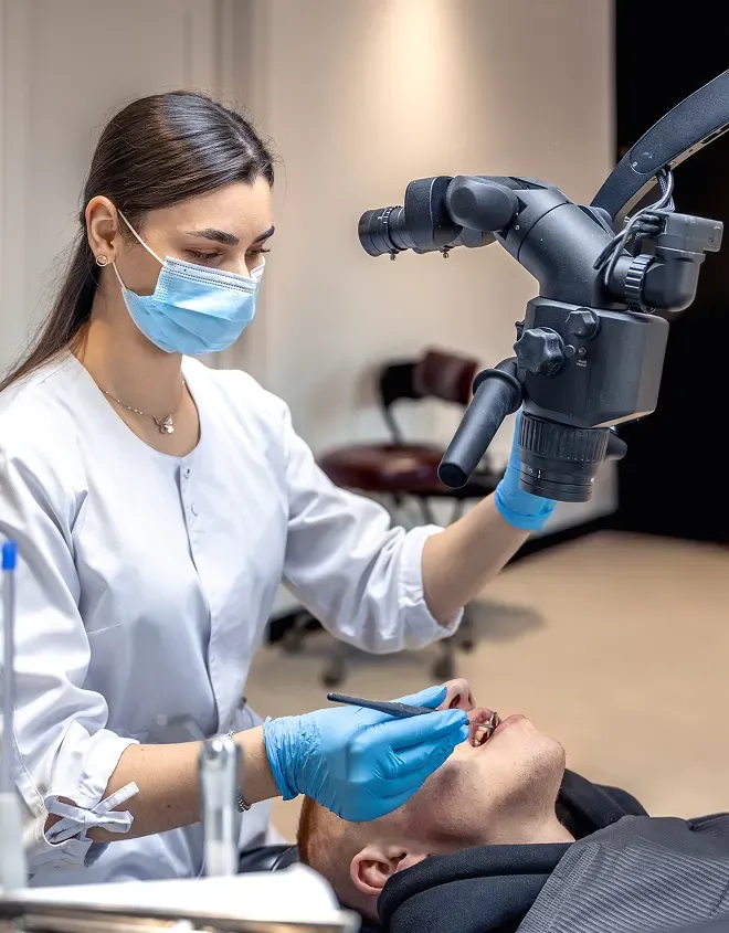
Intraoral 3D scanners play a pivotal role in transforming conventional oral surgery workflows. These scanners capture digital impressions for oral surgery that are highly accurate and immediately usable for surgical planning. The immediate availability of digital models allows dental teams to collaborate effectively, refine surgical guides, and simulate procedures prior to entering the operating room.
The precision achieved with these scans significantly improves outcomes in precision oral surgery. For example, in dental implant placement, a digitally planned procedure ensures that implants are positioned optimally within the jawbone. This meticulous planning reduces the likelihood of post-operative complications, enhances implant longevity, and improves overall patient satisfaction. Similarly, for facial reconstructive surgeries, the high-resolution 3D models facilitate exact alignment and fitting of grafts or prosthetics, a level of precision that was challenging to achieve with traditional methods.
Integration with Digital Surgical Planning Software
The real strength of 3D scanning lies not only in capturing detailed images but also in integrating these scans with digital surgical planning software. This software allows surgeons to manipulate the 3D models, simulate different approaches, and make adjustments without any risk to the patient. Combining this technology with 3D guided oral surgery Pinole techniques ensures that surgical procedures follow the pre-planned paths accurately.
Such integration is particularly valuable for complex procedures that involve multiple surgical sites or require simultaneous adjustments to facial structures. By relying on computer-assisted planning, surgeons can predict challenges, optimize the sequence of interventions, and enhance the overall precision of the procedure. Patients benefit from reduced operative times, minimized trauma, and improved recovery trajectories.
Moreover, the use of 3D digital scanning supports a more collaborative environment between oral surgeons and restorative dental professionals. The shared digital models enable teams to coordinate implant placements, prosthetic designs, and bone grafting procedures seamlessly. This collaboration ensures that the surgical outcomes align closely with the intended functional and aesthetic goals, demonstrating how high-precision facial surgery technology is now attainable in everyday clinical practice.
Current Trends in 3D Digital Scanning for Surgery
The adoption of 3D digital scanning is not just a technological upgrade, it reflects an ongoing trend in precision dentistry and surgery. More clinics in Pinole and across California are incorporating 3D dental scanning into their workflows to enhance patient outcomes. This trend emphasizes preventive planning, reduced invasiveness, and the ability to deliver highly customized surgical solutions.
Advanced imaging, combined with 3D guided surgical techniques, is setting a new standard in oral and facial procedures. Surgeons can now rely on data-driven insights rather than estimations, improving consistency and predictability across a wide range of treatments. Patients increasingly expect minimally invasive procedures with high accuracy, and digital scanning addresses both these expectations effectively.
Enhancing Surgical Accuracy with CBCT and 3D Scanning
The combination of CBCT (Cone Beam Computed Tomography) with intraoral 3D scanners provides an unprecedented level of detail for oral and facial surgery. Surgeons can now overlay CBCT data onto 3D digital scans, creating a precise anatomical map for precision oral surgery in Pinole. This fusion of imaging technologies allows for better evaluation of critical structures such as nerves and sinuses, ensuring that implants, grafts, or corrective procedures are placed with millimeter-level accuracy. The result is a safer surgical environment and improved patient confidence.
Benefits for Complex Facial Procedures
High-precision procedures, including orthognathic surgery, trauma reconstruction, and tumor removal, benefit significantly from high-precision facial surgery technology. 3D scanning enables surgeons to replicate facial anatomy accurately and plan interventions that align with both functional and aesthetic goals. Accurate surgical planning with 3D scans reduces guesswork during surgery, minimizes tissue trauma, and improves post-operative recovery. Surgeons can simulate different surgical approaches and predict outcomes, ensuring that the procedure is both efficient and effective.
Streamlining Workflow with Digital Surgical Planning Software
Integrating 3D scans into digital surgical planning software allows oral surgeons to convert data into actionable surgical guides. These guides direct the surgical instruments along precise trajectories, facilitating 3D guided oral surgery Pinole. This level of automation in planning ensures consistency across multiple procedures and reduces variability between operators. Patients benefit from shorter surgical times, fewer complications, and better functional and cosmetic outcomes.
Conclusion
The evolution of 3D digital scanning represents a significant leap forward in oral and facial surgery. By combining intraoral 3D scanners, CBCT imaging, and digital surgical planning software, surgeons can achieve unprecedented levels of precision. Patients experience safer, faster, and more predictable procedures, while clinicians benefit from enhanced planning and execution capabilities. As adoption continues and technology evolves, oral and facial surgery is entering a new era of precision, efficiency, and patient-centered care. For anyone seeking advanced surgical solutions, 3D digital scanning is not just a tool; it is a transformative approach that sets the standard for modern oral and facial surgery.
Disclaimer
*This media/content or any other on this website does not prescribe, recommend, or prevent any treatment or procedure. Therefore, we highly recommend that you get the advice of a qualified dentist or other medical practitioners regarding your specific dental condition. *